





Image information begins with the Source–Object–Detector geometry
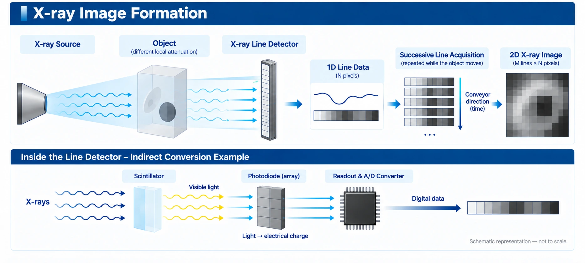
X-rays generated by the source are attenuated by different amounts as they pass through different parts of the object. Regions with different material composition, density, or thickness produce different transmitted intensities, and the detector placed behind the object measures these spatial differences in transmitted signal.
The most fundamental information in an X-ray image is therefore the position-dependent difference in X-ray intensity reaching the detector after passing through the object. Scatter reaching the detector, changes in the source spectrum, and inspection geometry can also affect the measured signal and image contrast.
The detector converts X-rays into digital signals
In a typical indirect-conversion X-ray detector, a scintillator converts part of the incident X-ray energy into visible light, and a photodiode converts that light into an electrical signal. The signal then passes through readout electronics and an A/D converter so that each pixel is represented as a digital value.
The conversion structure can differ depending on detector technology. For example, a direct-conversion detector collects electrical charge generated directly by X-rays without a separate scintillator. The overall principle of image formation is common, but the detailed process of converting X-rays into electrical signals depends on the detector technology.
How line-scan data become a two-dimensional image
An X-ray line detector acquires one-dimensional line data along the detector length in each acquisition. If the object moves at a constant speed and lines are acquired at regular intervals, each line corresponds to a different position along the direction of travel.
By stacking these line data in the order in which they are acquired, a two-dimensional X-ray image is formed: the horizontal direction represents the detector pixel array, while the vertical direction represents time and position as the object moves. Spatial sampling in the travel direction depends on the relationship between conveyor speed and line rate, so these two conditions must be set together.
Pixel values, image brightness, and contrast
The digital value of each pixel reflects the X-ray signal reaching the detector together with the detector response. In general, signal differences between regions of higher and lower attenuation provide the basis for image contrast. In a practical displayed image, however, calibration, gain / offset correction, normalization, and other image-processing steps may also be applied.
Whether a high transmitted signal is displayed as bright or dark can depend on the display convention or software settings. A bright or dark region in an X-ray image should therefore not be assigned a physical meaning without also considering the data-processing and display convention being used.
Key factors that determine image quality
Final X-ray image quality is not determined by transmission contrast alone. Source energy and output, object attenuation, detector pixel pitch and sensitivity, integration time, line rate, scattered radiation, and electronic noise can all contribute.
Detector calibration, bad-pixel correction, signal normalization, and post-processing can also affect image uniformity and visibility. An industrial inspection system should therefore be optimized as a complete system that includes the source, object, detector, mechanical conditions, and data-processing chain.
- The fundamental information in an X-ray image is the position-dependent difference in X-ray intensity reaching the detector after passing through the object.
- An indirect-conversion detector typically converts X-rays into digital pixel data through a scintillator, photodiode, readout electronics, and A/D conversion.
- A line detector acquires one-dimensional line data, and repeated lines acquired during motion are stacked in sequence to form a two-dimensional image.
- The bright / dark display direction can be reversed by software, so the display convention should be distinguished from the meaning of the raw signal.
- Image quality is influenced by the source, object, detector, scan conditions, calibration, and image processing together.
- IAEA — Diagnostic Radiology Physics: A Handbook for Teachers and Students, sections on digital X-ray imaging, image receptors and image quality.
- IAEA — Design, Development and Optimization of a Low Cost System for Digital Industrial Radiology, IAEA Radiation Technology Reports No. 2.


